Scientist, Robarts Research Institute Professor, Medical Biophysics, University of Western Ontario
“We’re looking to identify new biomarkers for early diagnosis and new targets for the treatment of Alzheimer’s disease by uncovering the earliest synaptic changes in the brain that lead to cell death.” Dr. Robert Bartha
When a cell is damaged by Alzheimer’s disease and the neurons in the brain begin to die, it can start a process of controlled cell death, known as apoptosis. Having a domino effect, once apoptosis begins it cannot be stopped. At least not yet.
Attempting to get ahead of this process, Dr. Robert Bartha leads a research team within the Canadian Consortium on Neurodegeneration in Aging (CCNA) who are developing methods to diagnose Alzheimer’s disease in its earliest stages and identify targets for treatment.
For this, they are looking at how cells communicate with each other through the synapses – i.e. the junction between two nerve cells in the nervous system.
Why the synapses?
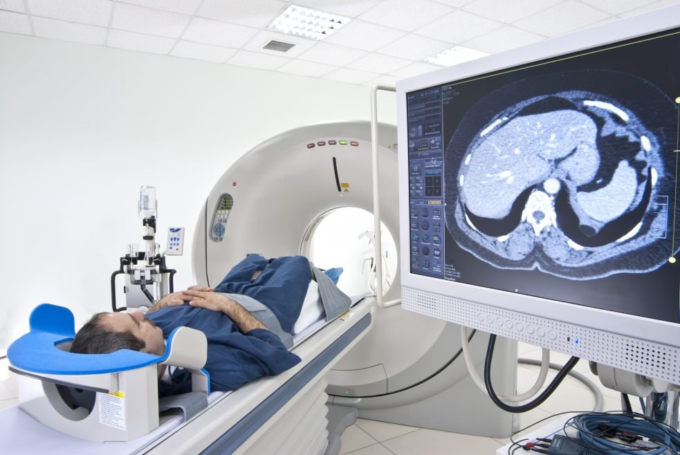
It’s true that researchers are already able to show when brain tissue is shrinking and dying by using MRI and PET-scans. Despite the valuable insights these imaging scans have brought Alzheimer’s disease researchers – enabling them to see amyloid plaques and tau deposits – this is already “later in the disease than we would like,” Bartha explains.
By successfully identifying the visual markers of apoptosis in the synapses, Bartha’s team could get at the “earliest damage in the brain before the bigger, more widespread changes occur.” In a word: They could stop the domino effect of apoptosis before it even begins.
To that end, they are building and validating a tracer that will target and illuminate a protein known as Caspase-3 that is associated with this cell death mechanism. In parallel, they’re creating a genetically-modified mouse model that will enable them to validate that the tracer is located where Caspase-3 is found within the mouse. Their end goal is to eventually use this technology to see Caspase-3, non-invasively, in humans.
To carry out this project, Bartha’s team had to build a foundation of new imaging techniques. This involved experimenting with a mouse brain (after the mouse was sacrificed) to dissolve away the lipids to effectively make the brain translucent. Researchers could then use Optical Imaging (a technique for non-invasively looking inside tissue) to show where Caspase-3 is located throughout the brain.
Taking a series of still images of what they saw, Bartha’s team converted those images into 3-D videos. Their goal is to use these imaging techniques to superimpose MRI and PET-scans of live mice on top of the translucent Optical Images and videos of the mouse to validate their new PET tracer. The idea is for them to map on – to show that the PET tracer is accumulating in the same place that the optical signal is changing. In this way, the team can verify that the imaging is actually measuring what they think it is measuring.
Collaborations and next steps
One of the team’s visions early on as part of the CCNA was for their work to funnel into Dr. Roger Dixon’s project that is developing new biomarkers. They intend to share what is being seen in mice and propose how Dixon’s team could apply their findings in, and use their novel imaging techniques for, humans.
Says Bartha, “if one of our projects works out really well, then within the CCNA we can potentially test it on a larger scale quickly in humans. And get results quickly.”
To learn more about Dr. Bartha and his team’s project, view their video by clicking here.